VesselWeaver
AbdVesselGen is an anatomy-guided abdominal vessel reconstruction framework for CT. It combines segmentation, anchor-point extraction, topology-aware pathfinding, and vessel graph generation to automatically trace major abdominal arteries and veins, with direct 3D Slicer SEG export support.
AbdVesselGen: Anatomy-Guided Abdominal Vessel Reconstruction for CT
AbdVesselGen is an anatomy-guided abdominal vessel reconstruction framework for contrast-enhanced abdominal CT.
The pipeline combines anatomical segmentation, anchor-point extraction, rule-guided vessel pathfinding, topology-aware post-processing, and automated vessel naming to reconstruct major abdominal arteries and veins. Final outputs can be exported as 3D Slicer-compatible .seg.nrrd segmentations for visualization, inspection, and downstream analysis.
Keywords
abdominal vessel reconstruction; abdominal CT angiography; vessel tracing; abdominal artery segmentation; portal vein segmentation; anatomy-guided pathfinding; topology-aware vessel reconstruction; 3D Slicer segmentation; TotalSegmentator; rule-based medical image processing; vascular graph reconstruction; abdominal vessel atlas.
📌 Overview
AbdVesselGen is designed to reconstruct and organize major abdominal vascular structures from CT using an interpretable, anatomy-guided workflow.
Unlike purely end-to-end deep learning segmentation pipelines, this project combines:
- TotalSegmentator-derived anatomical landmarks
- Vessel candidate segmentation
- Organ and hilum anchor-point extraction
- Anatomical plane generation
- HU-guided pathfinding
- Topology-aware vessel tracing
- Rule-based vessel naming
- 3D Slicer-compatible segmentation export
The goal is to create an inspectable abdominal vessel reconstruction pipeline that can help generate structured vascular maps from abdominal CT.
This project is designed for:
- Abdominal vessel tracing
- CT-based vascular atlas generation
- Radiology AI preprocessing
- Surgical anatomy visualization
- Rule-guided abdominal artery and vein reconstruction
- 3D Slicer-based review and correction workflows
🖼️ Demo
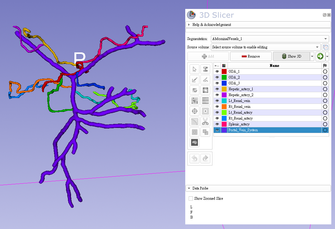
Abdominal vessel branch reconstruction
The following image shows an example output of the final abdominal vessel branch reconstruction in 3D Slicer.
🎥 YouTube demonstration:
https://youtu.be/9aVzKN6tO0s
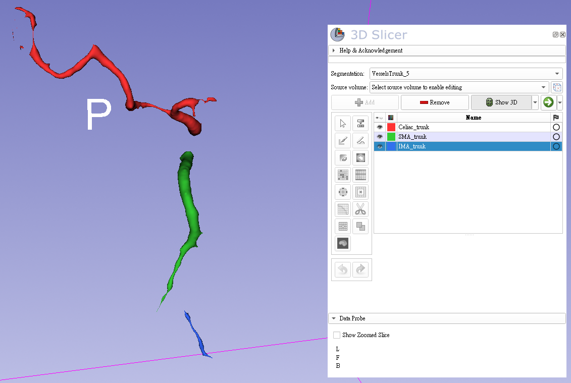
Abdominal vessel trunk reconstruction
The following image shows an example output of the reconstructed abdominal vessel trunks in 3D Slicer.
🎥 YouTube demonstration:
https://youtu.be/vPpqlPF7ano
🧠 Pipeline
Abdominal CT (.nii / .nii.gz)
↓
GUI-based case loading
↓
TotalSegmentator anatomical segmentation
↓
Tissue / cavity / liver segment extraction
↓
Colon vessel / portal venous candidate segmentation
↓
Anchor-point and anatomical plane generation
↓
Vessel start-point detection
↓
HU-guided vessel pathfinding
↓
Arterial and venous branch reconstruction
↓
Component naming and post-processing
↓
Export:

Intermediate masks (.nii.gz)
Named trunk segmentation (.seg.nrrd)
Named abdominal vessel branch segmentation (.seg.nrrd)
🩻 Target Vessels and Structures
The current pipeline focuses on major abdominal vascular structures, including:
Celiac trunk
SMA trunk
IMA trunk
Gastroduodenal artery
Hepatic artery branches
Splenic artery
Left renal vein
Right renal vein
Left renal artery
Right renal artery
Portal vein system
The framework is modular, so additional vessel branches can be added as independent scripts and inserted into the pipeline.
🧩 Core Concepts
1. Anatomy-guided reconstruction
The pipeline uses anatomical structures such as the aorta, IVC, liver segments, spleen, renal hilum, hepatic hilum, duodenum surface, and vertebral-level planes to define plausible vascular regions and endpoints.
2. Anchor-point based pathfinding
Instead of blindly tracing vessels from arbitrary seeds, the pipeline generates anatomical anchor points such as:
hepatic hilum spheres
spleen hilum point
renal hilum centroid spheres
duodenum surface anchors
renal artery / vein start candidates
search-volume derived starting points
These anchors constrain vessel pathfinding to anatomically meaningful start and end locations.
3. HU-guided vessel tracing
Pathfinding is performed on CT intensity space. Low-HU or invalid regions are penalized or excluded, while enhancing vascular voxels are preferred.
Typical forbidden or penalized regions include:
HU == -1024
outside ROI_CT
previously traced vessel paths
anatomically implausible regions
4. Independent segment layers
Final .seg.nrrd outputs store vessels as independent segment layers. This allows overlapping segments when anatomically or algorithmically necessary.
📁 Project Structure
AbdVesselGen/
├── run.py
│
├── Segmentator/
│   ├── AllSeg.py
│   ├── MoveAllSeg.py
│   ├── CavitySeg.py
│   ├── MoveCavitySeg.py
│   ├── TissueSeg.py
│   ├── MoveTissueSeg.py
│   ├── LiverSegmentsSeg.py
│   ├── MoveLiverSegmentsSeg.py
│   └── ColonVesselSeg/
│       ├── scripts/
│       │   ├── inference.py
│       │   ├── utils_inference.py
│       │   ├── prune.py
│       │   └── skeletonize.py
│       └── weights/
│           └── veins_segmentation_model.pth
│
├── Preprocess/
│   ├── Crop_ROI_CT.py
│   ├── FindRenalHilum.py
│   ├── FindSpleenHilum.py
│   ├── SearchStartCombined.py
│   ├── SearchStart_RenalA_RenalV.py
│   ├── RenalStartExtract.py
│   ├── FindAnchorPointsFromSearchVolumes.py
│   ├── FindDynamicCenterDuodenumSurface.py
│   └── GenerateDuodenumSurfaceAnchors.py
│
├── VesselsFinder/
│   ├── FindWeb_GDA.py
│   ├── Pathfind_HepaticArtery.py
│   ├── RenalPathfind.py
│   └── Pathfind_SplenicArtery.py
│
├── Postprocess/
│   ├── TrunkSeperator.py
│   └── BranchesCombine.py
│
├── Seg_Done/
│   ├── <case_id>_All_segmentation/
│   ├── <case_id>_trunk_cavities/
│   ├── <case_id>_tissue_4_types/
│   ├── <case_id>_liver_segments/
│   └── <case_id>_veins_segmentation/
│
├── ROI_CT/
│   └── <case_id>_ROI_CT.nii.gz
│
├── AnchorPoints/
│   ├── <case_id>_seg32_highest_seg31_lowest_Zplanes.nii.gz
│   ├── <case_id>_seg29_highest_seg28_lowest_Zplanes.nii.gz
│   ├── <case_id>_search3231_volume.nii.gz
│   ├── <case_id>_search2928_volume.nii.gz
│   ├── <case_id>_anchor_points_from_search_volumes.txt
│   ├── <case_id>_renal_hilum_seg2_seg3_centroid_spheres_diameter6.nii.gz
│   ├── <case_id>_SpleenHilum.nii
│   └── <case_id>_hepatic_hilum_spheres.nii
│
└── Results/
    ├── Intermediate/
    │   ├── <case_id>_GDA.nii.gz
    │   ├── <case_id>_hepatic_vessels.nii.gz
    │   ├── <case_id>_renal_artery_vein_paths.nii.gz
    │   └── <case_id>_splenic_artery_path.nii.gz
    │
    ├── VesselsTrunk.seg.nrrd
    ├── AbdominalVessels.seg.nrrd
    ├── <case_id>_VesselsTrunk_summary.txt
    └── <case_id>_AbdominalVessels_summary.txt
📂 Input Format
The pipeline currently supports a single CT NIfTI file selected through the GUI.
Supported formats:
.nii
.nii.gz
Example:
D:\rsna_bowel_injury\nifti_dataset\43_24055.nii
When loaded through run.py, the case ID is inferred from the CT filename:
43_24055.nii → case_id = 43_24055
All downstream outputs are automatically named using this case_id.
🚀 Usage
1. Install dependencies
Recommended environment:
conda create -n abdvesselgen python=3.10
conda activate abdvesselgen
Install core dependencies:
pip install numpy scipy nibabel pynrrd nilearn tqdm
Install medical segmentation dependencies as needed:
pip install torch monai simpleitk
pip install TotalSegmentator
If using the colon vessel segmentation module, place the pretrained model under:
Segmentator/ColonVesselSeg/weights/veins_segmentation_model.pth
2. Run the GUI pipeline
python run.py
Then:
- Select the abdominal CT NIfTI file
- Confirm the selected case
- Click Run Pipeline
- Monitor real-time logs in the GUI
- Review outputs in Results/
The GUI supports automatic step skipping if required outputs already exist.
📤 Output
1. ROI CT
ROI_CT/<case_id>_ROI_CT.nii.gz
This file contains the cropped / masked CT region used for downstream vessel pathfinding.
2. Anchor points
AnchorPoints/
Contains anatomical anchors and candidate search masks, such as:
<case_id>_search3231_volume.nii.gz
<case_id>_search2928_volume.nii.gz
<case_id>_anchor_points_from_search_volumes.txt
<case_id>_renal_hilum_seg2_seg3_centroid_spheres_diameter6.nii.gz
<case_id>_SpleenHilum.nii
<case_id>_hepatic_hilum_spheres.nii
3. Intermediate vessel masks
Results/Intermediate/
Typical outputs include:
<case_id>_GDA.nii.gz
<case_id>_hepatic_vessels.nii.gz
<case_id>_renal_artery_vein_paths.nii.gz
<case_id>_splenic_artery_path.nii.gz
4. Vessel trunk segmentation
Results/VesselsTrunk.seg.nrrd
This file contains named arterial trunk segments.
Naming rule:
If 3 components:
highest Z-axis component  = Celiac_trunk
middle Z-axis component   = SMA_trunk
lowest Z-axis component   = IMA_trunk

If 2 components:
highest Z-axis component  = Celiac+SMA_trunk
lowest Z-axis component   = IMA_trunk
5. Final abdominal vessel segmentation
Results/AbdominalVessels.seg.nrrd
This file combines major vessel branches into a 3D Slicer-compatible segmentation.
Segment naming rules:
GDA.nii.gz:
GDA_1, GDA_2, GDA_3, ...

hepatic_vessels.nii.gz:
Hepatic_artery_1, Hepatic_artery_2, ...

renal_artery_vein_paths.nii.gz:
Segment_1 → Lt_Renal_vein
Segment_2 → Rt_Renal_vein
Segment_3 → Lt_Renal_artery
Segment_4 → Rt_Renal_artery

splenic_artery_path.nii.gz:
Splenic_artery

portal venous segmentation:
Portal_Vein_System
Because each vessel is stored as an independent segment layer, overlapping segments are allowed.
🧠 Method Highlights
1. TotalSegmentator-based anatomical segmentation
The pipeline uses TotalSegmentator-derived anatomical labels as spatial priors.
Key structures may include:
aorta
inferior vena cava
liver
spleen
kidneys
vertebrae
trunk cavities
tissue classes
abdominal organs
These structures are used for ROI generation, anchor extraction, anatomical plane construction, and pathfinding constraints.
2. ROI CT generation
The pipeline creates an ROI CT by combining anatomical masks and excluding irrelevant or invalid regions.
The ROI CT is used as the main search space for pathfinding.
Invalid regions are typically encoded as:
HU = -1024
Pathfinding scripts avoid these regions.
3. Anatomical plane generation
Several scripts generate anatomical Z-plane constraints based on TotalSegmentator labels.
Examples:
seg32_highest_seg31_lowest_Zplanes
seg29_highest_seg28_lowest_Zplanes
These planes restrict where certain start-search algorithms are allowed to operate.
4. Vessel start-point detection
The pipeline searches for plausible vessel origins near major vascular structures.
Examples:
renal artery / vein candidate boxes
search3231_volume
search2928_volume
anchor_points_from_search_volumes.txt
Start points are selected using connected component centroids and validated against ROI CT intensity values.
5. Hilum and organ anchor extraction
The pipeline automatically extracts anatomical target points such as:
renal hilum centroid spheres
spleen hilum point
hepatic hilum spheres
duodenal internal surface anchors
These anchor points serve as vessel endpoints or intermediate constraints.
6. HU-guided pathfinding
Vessel branches are reconstructed using pathfinding over CT intensity space.
The pathfinding cost function generally:
penalizes low-HU regions
avoids HU == -1024
prefers enhancing vascular voxels
avoids previously traced vessel masks when needed
This allows the pipeline to trace plausible vessel routes between anatomical start and endpoint anchors.
7. Direction-constrained splenic artery tracing
For splenic artery pathfinding, an initial forward-only constraint can be used.
Example:
first 10 steps must move toward decreasing Y-axis value
This helps prevent the path from immediately traveling backward into incorrect high-HU regions.
8. Overlap-preserving Slicer export
Final .seg.nrrd outputs are written using independent segment layers:
(num_segments, X, Y, Z)
This preserves overlapping vessels and avoids accidental overwriting that can occur in single-label 3D labelmaps.
🖥️ GUI Features
The included Tkinter GUI supports:
manual CT NIfTI selection
automatic case ID extraction
one-click pipeline execution
real-time log display
progress-bar cleanup for subprocess logs
automatic output directory refresh
step skipping if output files already exist
total runtime measurement
This makes the pipeline easier to run repeatedly during development and debugging.
⚙️ Processing Time
Processing time depends on:
CT volume size
TotalSegmentator runtime
GPU availability
number of vessel pathfinding steps
whether intermediate outputs already exist
The pipeline prioritizes anatomical interpretability and reproducibility over raw speed. (It takes about 30 minutes for a case)
🔧 Adding New Pipeline Steps
Each pipeline step follows the same structure in run.py:
def step_new_module(ctx: PipelineContext) -> None:
    run_external_script(
        ctx,
        script_relative_path="Folder/NewModule.py",
    )
Optional skip check:
def should_skip_new_module(ctx: PipelineContext) -> bool:
    output_path = Path(
        rf"C:\Users\User\Desktop\AbdVesselGen\Results\Intermediate\{ctx.case_id}_new_output.nii.gz"
    )
    return output_path.exists()
Then insert into:
PIPELINE_STEPS = [
    ...
    PipelineStep(
        name="Step X - New Module",
        description="Description of the new module.",
        runner=step_new_module,
        enabled=True,
        skip_check=should_skip_new_module,
    ),
]
This modular structure allows new vessel branches, new anatomical anchors, and new post-processing steps to be added incrementally.
⚠️ Notes
This project is under active development. The current implementation is rule-based and anatomy-guided. It depends on the quality of upstream segmentation outputs. Poor contrast timing, motion artifact, unusual anatomy, or segmentation failure may affect vessel tracing. Manual inspection in 3D Slicer is recommended. Some scripts currently assume a Windows-style project path and may require path modification for other platforms.
⚠️ Disclaimer
This project is intended for research and educational purposes only.
It is not a certified medical device and should not be used as the sole basis for clinical diagnosis, treatment planning, operative planning, or clinical decision-making.
All outputs should be reviewed by qualified medical professionals before any research or clinical interpretation.
📄 License
MIT License
🙌 Acknowledgements
This project builds on or interacts with several open-source tools and medical imaging platforms:
TotalSegmentator

3D Slicer
MONAI
Nibabel
pynrrd
SciPy
NumPy
Nilearn
The portal vein system segmentation component of this project primarily uses the pretrained model and implementation provided by:
LERCO-FNO / Abdominal-Vessels-Segmentation
https://github.com/LERCO-FNO/Abdominal-Vessels-Segmentation
💡 Future Work
Planned or possible future improvements include:
batch processing support
cross-platform path handling
better anatomical variant handling
more robust vessel graph validation
automatic QA visualization
branch-level confidence scoring
semi-automatic manual correction tools
improved venous / arterial separation
quantitative vascular measurements
integration with radiology AI workflows
Citation
If you use this project in academic work, please cite this repository.
A formal citation will be added once a manuscript or preprint is available.